Research Interest
In humans, Plasmodium parasites cause malaria, a deadly disease killing hundreds of thousands of people each year. The sexual replication of Plasmodium, however, occurs in the mosquito, starting with the formation of male and female gametes (gametogenesis). Male gametogenesis is particularly fascinating, as within only ten minutes, eight male gametes form and emerge from one single parent cell. In this time the parent cell replicates its DNA three times to provide eight genomes, but its nucleus only divides when the eight gametes emerge from the cell. It is thus a challenge for the parasite to make sure that each gamete takes up a complete genome from the single nucleus. Failure to do so results in male gametes with too little DNA, and while they can still fertilise female gametes and form zygotes, these will then stop developing at the oocyst stage in the mosquito. A functional and complete male genome is thus essential for the parasite to transmit through the mosquito.

At the Hentzschel lab, we investigate the formation and contribution of the male genome to the sexual replication of Plasmodium. How does the male gametocyte ensure that each gamete takes up a complete genome? What does the male gamete contribute to the female gamete during fertilisation to initiate development of a motile zygote? How is replication organised in the oocyst and why does the oocyst require a diploid genome for this? We address these research questions using a combination of reverse genetics, live cell and fixed imaging techniques, and interactome studies to reveal the molecular basis for these phenotypes. In addition, we develop novel genetic tools to enable investigation of gene function in the diploid oocyst stages.
Lab members
Current members
Dr. Franziska Hentzschel (Group leader, 2023 – present)
Lukas Baltes (PhD student, 2024)
Katharina Zorko (MSc thesis, 2025)
Nora Langner (BSc Thesis, 2024; HiWi, 2025)
Maximilian Vassen (BSc Thesis, 2025)
Alumni
Viola Reuschenbach (BSc internship, 2024)
Verena Kantelhardt (BSc Thesis, 2024)
Anna Kraeft (Internship, 2024)
Lukas Brenner (MSc internship, 2022; MSc Thesis, 2023-2024)
Fenja Nuglisch (Internship, 2024)
David Lubotsky (MD Thesis, 2022 - 2024)
Yvonne Sokolowski (MSc Thesis, 2023)
David Jewanski (BSc Thesis, 2023)
Pratika Agarwal (MSc Thesis, 2022 – 2023)
Projects
Deciphering DNA segregation during Plasmodium male gametogenesis

During the rapid formation of male gametes, the cell must ensure that each of the eight budding gametes takes up one genome from the octoploid nucleus. We have discovered a protein complex that is essential for this DNA segregation. We are now investigating its mechanistic function and how this protein complex is assembled and activated. We also investigate further interaction partners to gain a deeper understanding of the molecular biology of male gamete formation.
Developing novel CRISPR tools to dissect the mosquito stages of Plasmodium

The molecular and genetic basis of the development of Plasmodium in mosquitoes is little understood. One reason for this is that genetic modifications can only be done in blood stages, and if a gene deletion arrests parasite development there, the function of this gene cannot be investigated in subsequent oocyst stages. Another reason is that oocysts are diploid, carrying both the maternal and the paternal genome, which further complicates reverse genetics. We are working on engineering CRISPR-based gene targeting strategies that make use of the sexual replication of Plasmodium to silence gene expression only in the diploid oocyst.
Investigating the early replication events in the oocyst

The oocyst is a highly unusual, underexplored life cycle stage of Plasmodium. Not only is it the only stage at which the parasite grows extracellularly, it also employs an unusual mode of replication where DNA replication is not always followed by nuclear division, and the oocyst thus contains multiple nuclei with several genomes each. Curiously, we found that if oocysts are aneuploid, i.e. missing a subset of the paternal genome, oocysts arrest early in development. Using live-cell imaging approaches, single-cell transcriptomics and reverse genetics, we aim to spatiotemporally characterize early oocyst development and to understand how the paternal genome is important for this development.
-

We always welcome applications from motivated students for lab rotations (min. 6 weeks) or BSc/MSc theses. Please send an informal inquiry including a CV & a short statement on your motivation via email.
Publications
Original articles
2023
Ferreira, J. L., Pražák, V., Vasishtan, D., Siggel, M., Hentzschel, F., Binder, A. M., Pietsch, E., Kosinski, J., Frischknecht, F., Gilberger, T. W., & Grünewald, K. (2023). Variable microtubule architecture in the malaria parasite. Nature Communications, 14(1), 1–17. DOI: 10.1038/s41467-023-36627-5
2022
Ripp, J., Smyrnakou, X., Neuhoff, M.-T., Hentzschel, F., & Frischknecht, F. (2022). Phosphorylation of myosin A regulates gliding motility and is essential for Plasmodium transmission. EMBO Rep, e54857. DOI: 10.15252/EMBR.202254857
Hentzschel, F., Gibbins, M. P., Attipa, C., Beraldi, D., Moxon, C. A., Otto, T. D., & Marti, M. (2022). Host cell maturation modulates parasite invasion and sexual differentiation in Plasmodium berghei. Sci Adv, 8(17), 7348. DOI: 10.1126/SCIADV.ABM7348
2021
Girard, A., Cooper, A., Mabbott, S., Bradley, B., Asiala, S., Jamieson, L., Clucas, C., Capewell, P., Marchesi, F., Gibbins, M. P., Hentzschel, F., Marti, M., Quintana, J. F., Garside, P., Faulds, K., MacLeod, A., & Graham, D. (2021). Raman spectroscopic analysis of skin as a diagnostic tool for Human African Trypanosomiasis. PLOS Pathogens, 17(11), e1010060. DOI: 10.1371/JOURNAL.PPAT.1010060
2020
Fernandez-Becerra, C., Bernabeu, M., Castellanos, A., Correa, B. R., Obadia, T., Ramirez, M., Rui, E., Hentzschel, F., … del Portillo, H. A. (2020). Plasmodium vivax spleen-dependent genes encode antigens associated with cytoadhesion and clinical protection. PNAS, 117(23), 13056–13065. DOI: 10.1073/pnas.1920596117
Hentzschel, F., Obrova, K., & Marti, M. (2020). No evidence for Ago2 translocation from the host erythrocyte into the Plasmodium parasite. Wellcome Open Res, 5, 92. DOI: 10.12688/wellcomeopenres.15852.2
Hentzschel, F., Mitesser, V., Fraschka, S., Krzikalla, D., Carrillo, E., Berkhout B., Bártfai R., Mueller, A.-K., & Grimm, D. (2020) Gene knockdown in malaria parasites via non-canonical RNAi. NAR, 48(1), e2. DOI: 10.1093/nar/gkz927
2014
Hentzschel, F.*, Hammerschmidt-Kamper, C.*, Börner, K.*, Heiss, K.*, (...), Mueller, A-K. M., Grimm, D. (2014) AAV8-mediated in vivo overexpression of miR-155 enhances the protective capacity of genetically-attenuated malarial parasites. Mol. Ther., 22(12), 2130–2141. DOI: 10.1038/mt.2014.172
(*: equal contribution)
Reviews
2022
Hentzschel, F., & Frischknecht, F. (2022). Still enigmatic: Plasmodium oocysts 125 years after their discovery. Trends in Parasitology, 38(8), 610–613. DOI: 10.1016/J.PT.2022.05.013 (Review)
2020
Venugopal, K., Hentzschel, F., Valkiūnas, G., Marti, M. (2020) Plasmodium asexual growth and sexual development in the haematopoietic niche of the host. Nat Rev Micro, 18(3), 177–189. DOI: 10.1038/s41579-019-0306-2 (Review)
2019
Ngotho P., Soares AB., Hentzschel F., Achcar F., Bertuccini L., Marti M. (2019) Revisiting gametocyte biology in malaria parasites. FEMS Microbiol Rev., 43(4), 401-414. DOI: 10.1093/femsre/fuz010 (Review)
2016
Hentzschel, F.*, Herrmann, A.-K., Mueller, A.-K.*, & Grimm, D. (2016) Plasmodium meets AAV - the (un)likely marriage of parasitology and virology, and how to make the match. FEBS Letters, 590(13), 2027–45. DOI: 10.1002/1873-3468.12187 (Review)
(*: equal contribution)
Preprints
Hentzschel, F., Jewanski, D., Sokolowski, Y., Agarwal, P., Kraeft, A., Hildenbrand, K., Dorner, L.P., Singer, M., Frischknecht, F., Marti, M. A non-canonical Arp2/3 complex is essential for Plasmodium DNA segregation and transmission of malaria. BioRxiv (Preprint)
Hentzschel, F., Binder, A.M., Dorner, L.P., Herzel, L., Nuglisch, F., Sema, M., Aguirre-Botero, M.C., Cyrklaff, M., Funaya, C., Frischknecht, F. Microtubule inner proteins of Plasmodium are essential for transmission of malaria parasites. BioRxiv (Preprint)
News - Hentzschel group

Rudolphi Medal for F. Hentzschel
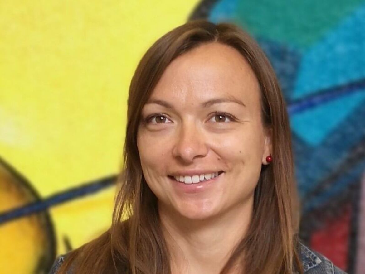
We are delighted to announce that our group leader Dr Franziska Hentzschel is the winner of this year's Karl Asmund Rudolphi Medal, awarded by the German Society of Parasitology (DGP). The medal was presented to her for her outstanding scientific achievements in the field of parasitology at the joint spring conference of the DGP, the British Society of Parasitology and the Swiss Society of Tropical Medicine and Parasitology, which took place in Würzburg from 11-14 March.
Congratulations!
ERC grant for F. Hentzschel
We are delighted to announce that our research group leader Dr Franziska Hentzschel has been awarded one of the prestigious and highly endowed Starting Grants from the European Research Council (ERC). This makes her the only scientist in Heidelberg to have achieved this in the current funding round. The 5-year grant of approximately 1.5 Mio Euros will be used to explore new strategies against malaria. We wish our research group leader every success in this endeavour!
