Pattern Analysis and Learning Group
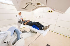
The field of digital oncology has brought the large-scale introduction of data science and artificial intelligence into the health care sector and cancer patient care. Today's advanced radiotherapy methods involve an enormous wealth of multimodal medical patient and especially image data, requiring the careful use of computer science to efficiently manage and facilitate information for the best treatment options. Artificial intelligence and deep learning technologies are applied to advance the current state of the art in radiooncology towards better assessment of diagnosis, risk and prognosis as well as planning and delivery of accurate treatments.
The section of “Pattern Analysis and Learning” pioneers research in machine learning and information processing, with the particular aim of improving cancer patient care by systematic data analytics. The section pursues cutting-edge developments at the core of computer science with applications in radiation oncology and is particularly interested in techniques for semi- and unsupervised learning and probabilistic modeling.
Our vision is to advance the quality of radiation oncology through methodological advances in artificial intelligence research and their large-scale clinical implementation. We therefore have a particular interest in techniques that improve the applicability of data science in clinical settings, e.g. by providing more interpretable decision-making, by explicitly dealing with data uncertainty, by increasing the generalizability of algorithms or by learning more powerful representations. We further study image computing concepts that combine mathematical modelling approaches with current machine learning techniques.
Relevant Projects
HiGHMed
HiGHmed is a highly innovative consortial project in the context of the “Medical Informatics Initiative Germany” that develops novel, interoperable solutions in medical informatics with the aim to make medical patient data accessible for clinical research in order to improve both, clinical research and patient care. Our image analysis technology (d:cipher) is part of the Omics Data Integration Center (OmicsDIC) that offers sophisticated technologies to process data and to access information contained in data - from genomics to radiomics. In HiGHmed we also improve the interoperability of image based information by working on the mapping between different important standards like DICOM, HL7 FHIR or OpenEHR.
Radiomics- and Shape-based Detection of Diabetes related Tissue Changes in the German National Cohort
This project aims at an extensive computational analysis of medical data from the multi-center study ‘German National Cohort (GNC)’. The GNC offers highly standardized MR image acquisition, holistic documentation of clinical parameters and patient history, as well as large case numbers organized in a nation-wide effort. This makes the examined GNC data an ideal subject for analysis using modern, data-driven machine-learning algorithms. Pre-diabetes and diabetes type 2 are likely associated with quantity and distribution of abdominal tissue types (adipose fat, or fat and iron in the liver) and with liver shape. In this project, information on tissue and shape characteristics will automatically be derived from GNC images and be correlated with pathologic findings from the GNC. The project will focus on strategies for computational quality-assessment of learning-based annotation algorithms, on continuous algorithm re-training, and on complementary radiomics analyses based on hand-crafted and deep learning derived image features.
End-to-end Deep learning Architecture
Current Medical Image Analysis approaches often comprise a set of separate processing steps such as Registration, Normalization, Segmentation, Feature Extraction and Classification. This project develops techniques for the integration of these components into one end-to-end deep learning architecture. This enables simultaneous optimization of all component w.r.t. the ultimate clinical task (e.g. disease classification).
Knowledge-based large-scale image segmentation
This project aims at the development of flexible and accurate methods for an automatic semantic image segmentation on large clinical datasets. We focus on a combination of model-based methods with techniques from Active Learning, Online Learning, Transfer Learning and Deep Learning. They allow an optimized training on sparse data, a continuous learning at runtime and an assessment of segmentation quality, as a prerequisite for the successful annotation of large and heterogeneous datasets.
Radiomics
In Radiomics, high-throughput computing techniques are systematically employed for the conversion of images to higher-dimensional data, i.e. predictive features. The aim is to improve decision support by the subsequent analysis of these features. We study comprehensive MRI imaging phenotypes for linkage of imaging with clinical, biological and genomic parameters in several entities including prostate cancer, breast cancer and brain tumors.
Image-based Stratification of Glioblastoma Patients
This project focuses on uncertainty in radiomics. Traditionally, a multitude of parameters is computed from images with which decision support systems are learned. These parameters may however be very sensitive to segmentation errors, resulting in overfitting and degrading performance. We make use of deep learning segmentation algorithms that allow uncertainty estimation, based upon which we can learn more robust decision support algorithms.
Longitudinal Modeling for Glioblastoma Assessment
In this project, we develop algorithms to predict future clinical assessments of glioblastoma patients from existing longitudinal data. We specifically try to estimate spatial invasion probabilities, incorporating novel uncertainty measures that allow us to quantify the confidence of our models’ outputs. This project is carried out in close cooperation with the Department of Neuroradiology at Heidelberg University Hospital, where we have established infrastructure that allows us to test cutting edge algorithms in clinical routine.
MITK Workbench
The MITK Workbench is a free medical image viewer, which supports all kinds of DICOM modalities like CT, MRT, US and RT as well as a number of research file formats. In addition to 2D, 3D and 3D+t visualization capabilities, there are numerous plugins for segmentation, registration and other processing steps available.
Research data management and automated processing
Additionally to the interactive processing focused in MITK, today's research questions often require the standardized and automated processing and easy data access while reducing emerging hassles of data transfer, data protection and data storage. We provide scientific cloud and platform solutions and evaluate new exploration and processing capabilities to medical imaging researchers.
RTT toolbox
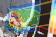
"RTToolbox" is a robust and flexible C++ software library developed to support quantitative analysis of treatment outcome for radiotherapy. It provides the import of radiotherapy data (e.g. plans, dose distributions and structure sets) in DICOM-RT standard format as well as in ITK supported data formats. Core features of the RTToolbox include: calculation of DVHs and dose comparison indices, and computation of radiobiological models (TCP/NTCP). Using the RTToolbox radiotherapy evaluation applications can be built easily and quickly.
Group members:
Ralf Floca, Dr. sc. hum.
Marco Nolden, Dr. rsc. hum.
Tobias Norajitra; Dr. ing.
Jan Sellner, MSc
Research opportunities:
Student Research Projects, Master Theses and PhD Dissertations
Are you interested in our research and would like to carry out a student research project, bachelor, master, or diploma thesis? Or would you like to write your PhD thesis in our division? We have changing funding and open positions. Thus, we are unable to maintain an updated list here.
Please send us an e-mail (mic-jobs(at)dkfz-heidelberg.de) with your application, providing a single pdf file containing the following:
- A motivational letter describing why you want to work at MIC or on a specific project and how your experience/education relates to this project
- A CV (limit two pages) including an overview of your programming skills
- A recent overview of grades from your studies, include final grades if possible
- A copy of your school graduation certificate
- Letters of references (if any)
