Onkologische Bildgebung in der RadioOnkologie und Strahlentherapie


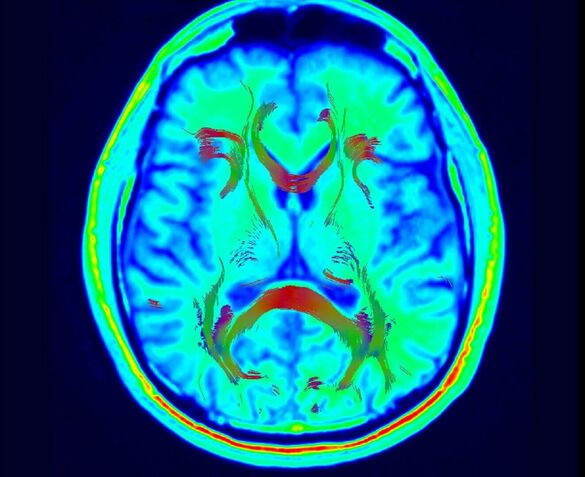
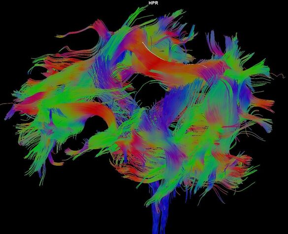
Die Forschungsgruppe Onkologische Bildgebung in der RadioOnkologie und Strahlentherapie konzentriert sich auf die Integration moderner bildgebender Verfahren in die strahlentherapeutische Behandlung von onkologischen Patienten. Ein Schwerpunkt liegt auf der Anwendung von MRT-basierten funktionellen Bildgebungstechniken im klinischen Alltag.
Das Spektrum reicht von tierexperimentellen präklinischen Studien, über Untersuchungen zur MR-Integration in die Bestrahlungsplanung bis hin zur Erfassung von Spätnebenwirkungen nach der Radiotherapie.
Oncologic Imaging in Radiotherapy
The research group Oncologic Imaging in Radiotherapy focused on the integration of modern imaging techniques, such as magnetic resonance imaging, into the radiotherapeutic treatment of oncological patients. Emphasis is placed on the application of MRI-based functional imaging techniques to clinical practice.
The spectrum of MRI examinations ranges from animal-experimental preclinical studies, to studies of MR integration in radiation planning and the recording of late side effects in radiotherapy.
Mitarbeiter/-innen
Kooperationspartner/-innen
Prof. Dr. Christian Karger (DKFZ- Abteilung Med. Physik in der Strahlentherapie)
Prof. Dr. Klaus Maier-Hein (DKFZ-Division of Medical Image Computing)
Prof. Dr. Frederike Rosenberger (NCT-Sportwissenschaft)
Forschungsschwerpunkte / Aktuelle Projekte
- Diffusionsbildgebung der Chordome und Chondrosarkome
- Diffusionsbildgebung zerebraler Nebenwirkungen nach Radiotherapie
- Tierexperimentelle MRT-Bildgebung zur Bestimmung der Nebenwirkungen nach Schwerionenionen-Radiotherapie [Kooperationspartner Prof. Dr. Christian Karger (DKFZ)]
- 3D-SPACE Bildgebung vor stereotaktischer Radiotherapie
Klinische Studien
- ARTEMIS
- SPACE-RT-Study
- CYBER-CHALLENGE
- CYBER-SPACE
- HIT-1-CHORDOM-STUDIE
- CS.pP.12C. CHONDROSARKOM-STUDIE
- ISAC –CHORDOM-STUDIE
- IRON II
Literatur / Publikationen
Three-dimensional SPACE imaging for MRI-based treatment planning (SPACE-RT-Study)
Welzel T, El Shafie RA, V Nettelbladt B, Bernhardt D, Rieken S, Debus J. Stereotactic radiotherapy of brain metastases: clinical impact of three-dimensional SPACE imaging for 3T-MRI-based treatment planning. Strahlenther Onkol. 2022 Oct;198(10):926-933. doi: 10.1007/s00066-022-01996-1.
MRI studies after carbon ion and photon irradiation
Bendinger AL, Welzel T, Huang L, Babushkina I, Peschke P, Debus J, Glowa C, Karger CP, Saager M. DCE-MRI detected vascular permeability changes in the rat spinal cord do not explain shorter latency times for paresis after carbon ions relative to photons.
Radiother Oncol. 2021 Dec; 165:126-134. doi: 10.1016/j.radonc.2021.09.035. Epub 2021 Oct 8.
Welzel T, Bendinger AL, Glowa C, Babushkina I, Jugold M, Peschke P, Debus J, Karger CP, Saager M. Longitudinal MRI study after carbon ion and photon irradiation: shorter latency time for myelopathy is not associated with differential morphological changes. Radiat Oncol. 2021 Mar 31;16(1):63. doi: 10.1186/s13014-021-01792-8.
Saager M, Peschke P, Welzel T, Huang L, Brons S, Grün R, Scholz M, Debus J, Karger CP. Late normal tissue response in the rat spinal cord after carbon ion irradiation. Radiat Oncol. 2018 Jan 11;13(1):5. doi: 10.1186/s13014-017-0950-5.
White matter changes after whole brain irradiation (Research granded by DFG)
Welzel T, Niethammer A, Mende U, et al. Diffusion tensor imaging screening of radiation-induced changes in the white matter after prophylactic cranial irradiation of patients with small cell lung cancer: first results of a prospective study. AJNR Am J Neuroradiol. 2008;29(2):379-383. doi:10.3174/ajnr. A0797
MR-Imaging of Chordomas and Chondrosarcomas
Welzel T, Meyerhof E, Uhl M, et al. Diagnostic accuracy of DW MR imaging in the differentiation of chordomas and chondrosarcomas of the skull base: A 3.0-T MRI study of 105 cases. Eur J Radiol. 2018; 105:119-124. doi:10.1016/j.ejrad.2018.05.026
Bostel T, Nicolay NH, Welzel T, Bruckner T, Mattke M, Akbaba S, Sprave T, Debus J, Uhl M. Sacral insufficiency fractures after high-dose carbon-ion based radiotherapy of sacral chordomas. Radiat Oncol. 2018 Aug 23;13(1):154. doi: 10.1186/s13014-018-1095-x.
Uhl M, Welzel T, Jensen A, Ellerbrock M, Haberer T, Jäkel O, Herfarth K, Debus J. Carbon ion beam treatment in patients with primary and recurrent sacrococcygeal chordoma. Strahlenther Onkol. 2015 Jul;191(7):597-603. doi: 10.1007/s00066-015-0825-3. Epub 2015 Mar 4.PMID: 25737378
Uhl M, Welzel T, Oelmann J, Habl G, Hauswald H, Jensen A, Ellerbrock M, Debus J, Herfarth K. Active raster scanning with carbon ions: reirradiation in patients with recurrent skull base chordomas and chondrosarcomas. Strahlenther Onkol. 2014 Jul;190(7):686-91. doi: 10.1007/s00066-014-0608-2. Epub 2014 Mar 25.
Uhl M, Mattke M, Welzel T, Roeder F, Oelmann J, Habl G, Jensen A, Ellerbrock M, Jäkel O, Haberer T, Herfarth K, Debus J. Highly effective treatment of skull base chordoma with carbon ion irradiation using a raster scan technique in 155 patients: first long-term results. Cancer. 2014 Nov 1;120(21):3410-7. doi: 10.1002/cncr.28877. Epub 2014 Jun 19
Side effects after Radiotherapy
Welzel T, Tanner JM. Nebenwirkungen nach Strahlentherapie in der Bildgebung [Imaging of side effects after radiation therapy]. Radiologe. 2018;58(8):754-761. doi:10.1007/s00117-018-0412-6
Bone Tumor Research Group
Rief H, Petersen LC, Omlor G, Akbar M, Bruckner T, Rieken S, Haefner MF, Schlampp I, Förster R, Debus J, Welzel T. The effect of resistance training during radiotherapy on spinal bone metastases in cancer patients - a randomized trial. Radiother Oncol. 2014;112(1):133-139. doi:10.1016/ j.radonc.2014.06.008


